

Introduction
A small pearly bump on your nose or cheek is easy to ignore. It may bleed once, scab over, and seem to heal. Then it returns. Many people shrug off such spots, especially after years of dacha summers and beach holidays in the south. However, this pattern can signal basal cell carcinoma, the most common type of skin cancer worldwide.
The good news is that basal cell carcinoma grows slowly and rarely spreads to other organs. Doctors can treat most cases successfully when they catch them early. In this guide, you will learn the warning signs, causes, diagnosis steps, treatment options, and prevention habits. Additionally, you will see when to book a dermatologist visit. Read on to protect your skin with facts, not fear.
What Is Basal Cell Carcinoma?
Basal cell carcinoma (BCC) is a cancer that starts in the basal cells. These cells sit in the deepest layer of the epidermis, the outer layer of your skin. Normally, they renew your skin by making new cells. However, DNA damage can make them multiply without control. The result is a growth that slowly forms a tumor.
Where Basal Cell Carcinoma Starts
Most tumors develop on skin that gets the most sun. Therefore, the face, ears, neck, scalp, and shoulders are common sites. Tumors can also appear on the chest and back. Basal cells exist in hair follicles too, so hairy areas are not immune.
How Common Is Basal Cell Carcinoma?
Dermatology organizations describe BCC as the most frequent skin cancer in fair-skinned people. Risk rises with age. However, studies suggest it increasingly appears in adults in their 30s and 40s, often after years of tanning.
Is Basal Cell Carcinoma Dangerous?
Basal cell carcinoma rarely spreads to distant organs. Nevertheless, it can invade nearby tissue, including cartilage and bone, when left untreated. Facial tumors may cause scarring or lasting damage. Additionally, small tumors need smaller surgery. Early action protects both your health and your appearance.
Basal Cell Carcinoma Symptoms: What Does It Look Like?
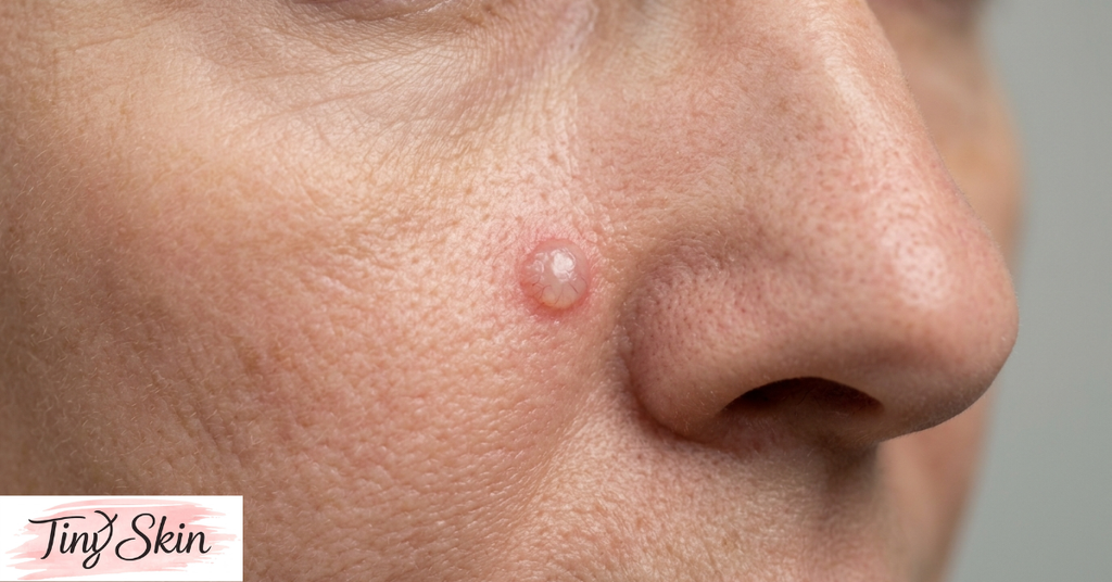
Basal cell carcinoma symptoms vary, so the disease is easy to miss. Most lesions look harmless at first. Dermatologists advise you to watch any spot that changes, bleeds, or refuses to heal.
Common Warning Signs
- A pearly, waxy, or flesh-colored bump
- Tiny visible blood vessels on the surface
- A sore that bleeds, scabs, and returns
- A flat, scaly pink or red patch
- A white or yellow scar-like area
- A bump with a rolled border or central dip
Where Basal Cell Carcinoma Appears
Sun-exposed spots such as the nose, eyelids, ears, and scalp are frequent targets. However, tumors can also develop on covered areas, such as the trunk. Therefore, check your whole body, not only your face.
When to See a Doctor
Book a visit if a spot grows, bleeds, itches, or does not heal within four weeks. Moreover, photograph suspicious spots monthly. Photos help your doctor judge change over time. Do not wait for pain, because these tumors are often painless. Additionally, mention any spot that a family member points out on your back or scalp, since you cannot see those areas easily.
Types of Basal Cell Carcinoma
Doctors group basal cell carcinoma by its pattern under the microscope and its look on the skin. The subtype affects treatment choice and recurrence risk.
Common Subtypes
| Subtype | Typical Appearance | Common Location | Key Note |
|---|---|---|---|
| Nodular | Pearly dome with visible vessels | Face | Most common subtype |
| Superficial | Flat, red, scaly patch | Trunk, shoulders | Often mistaken for eczema |
| Morpheaform (sclerosing) | Waxy, scar-like plaque | Face | Hard to see; may need Mohs surgery |
| Pigmented | Brown or blue-black bump | Sun-exposed skin | Can mimic melanoma |
| Infiltrative | Poorly defined edges | Face, scalp | Grows deeper; higher recurrence risk |
Why the Subtype Matters
Nodular and superficial types usually respond well to standard treatment. In contrast, morpheaform and infiltrative types spread under the skin surface with unclear borders. Surgeons may then miss tumor edges. Consequently, doctors often choose Mohs surgery for these tougher cases. Your biopsy report names your subtype, so ask your doctor to explain it.
Causes and Risk Factors for Basal Cell Carcinoma
Several factors raise your risk. You can control some of them, but not others.
Ultraviolet Radiation
Ultraviolet (UV) radiation is the leading cause. Sunlight and tanning beds both damage DNA in skin cells. Researchers link intense, intermittent sun exposure and childhood sunburns to basal cell carcinoma. Therefore, a sudden beach holiday after a long, dark winter can harm unprepared skin.
Personal Risk Factors
- Fair skin, light eyes, or red or blond hair
- A history of sunburns or regular tanning
- Age over 50
- A previous skin cancer
- A family history of skin cancer
- A weakened immune system, such as after an organ transplant
- Gorlin syndrome, a rare inherited condition
Other Triggers
Past radiation therapy, long-term arsenic exposure, and old scars or chronic wounds can also play a role. Furthermore, people who work outdoors, such as builders, farmers, and fishermen, face higher UV exposure over decades. If you fall into this group, schedule regular skin exams even when you feel well.
How Doctors Diagnose Basal Cell Carcinoma
An accurate diagnosis starts with a careful look at your skin. Fortunately, the process is quick and low-risk.
Skin Exam and Dermoscopy
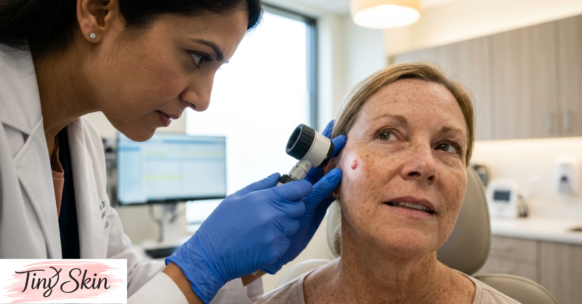
Your dermatologist examines your skin from head to toe. Next, they use a dermatoscope, a handheld magnifier with built-in light. It reveals vessel patterns and structures that the naked eye cannot see. This step helps separate harmless spots from suspicious ones.
Biopsy Confirms the Diagnosis
A biopsy confirms basal cell carcinoma. The doctor numbs the area and removes a small sample. A pathologist then studies it under a microscope. Results usually take several days to two weeks. The report also names the subtype and tumor depth.
Conditions That Look Similar
Several skin problems mimic BCC:
- Squamous cell carcinoma, another common skin cancer
- Melanoma, especially when compared with pigmented tumors
- Actinic keratosis, a rough, precancerous sun-damage patch
- Eczema or psoriasis, which resemble superficial tumors
- Benign moles and cysts
Because of these look-alikes, never self-diagnose. Only a doctor can tell them apart.
Basal Cell Carcinoma Treatment Options
Treatment depends on the subtype, size, location, and your overall health. Fortunately, most options work well when you start early. A dermatologist, surgeon, or oncologist will recommend a plan.
| Treatment | Best For | How It Works | Consideration |
|---|---|---|---|
| Standard excision | Most small to medium tumors | Removes tumor plus a safety margin | Leaves a scar; lab checks margins |
| Mohs surgery | Face, recurrent or aggressive tumors | Removes thin layers, checked immediately | Highest cure rates; saves healthy tissue |
| Curettage and electrodesiccation | Small, low-risk tumors | Scrapes and cauterizes the tumor | Quick; may leave a pale scar |
| Cryotherapy | Small, superficial tumors | Freezes tissue with liquid nitrogen | Causes blistering; no margin check |
| Imiquimod or 5-fluorouracil cream | Superficial tumors | Applied for several weeks | Causes redness and irritation |
| Radiation therapy | Patients unfit for surgery | Targeted beams destroy cells | Needs multiple sessions |
| Hedgehog inhibitors | Advanced tumors | Oral pills block growth signals | Side effects need monitoring |
Surgical Options
Surgery is the most common approach. Standard excision removes the tumor with a small margin of healthy skin. Mohs surgery removes thin layers and checks each one under the microscope right away. Studies suggest Mohs offers the highest cure rates for facial and recurrent tumors. However, availability varies by region, so ask your clinic.
Non-Surgical Options
Small, low-risk tumors may respond to curettage, freezing, or medicated creams. Radiation suits patients who cannot have surgery. Importantly, avoid folk remedies such as celandine juice or herbal pastes. They can burn healthy skin and delay real treatment.
Advanced Basal Cell Carcinoma
Rarely, tumors grow large or invade deeply. In these cases, specialists may prescribe hedgehog pathway inhibitors, oral medicines that block tumor growth signals. Your care team will monitor side effects closely.
How to Prevent Basal Cell Carcinoma
Prevention works better than any cure. Dermatologists recommend a few simple habits.
Smart Sun Protection
- Apply broad-spectrum SPF 30+ sunscreen every morning on exposed skin.
- Reapply every two hours outdoors and after swimming.
- Seek shade between 11:00 and 16:00.
- Wear a wide-brim hat, sunglasses, and long sleeves.
- Skip tanning beds completely.
Regular Skin Checks
Examine your skin monthly in good light. Use a mirror for your back and scalp. Moreover, see a dermatologist yearly if you have fair skin, many moles, or past skin cancer. Early detection keeps treatment simple.
Protection All Year in Russia
UV rays pass through clouds. Snow also reflects them, so ski trips and winter fishing need sunscreen too. Before southern holidays on the Black Sea coast or abroad, build protection with shade and clothing instead of fast tanning. Gradual, careful exposure beats painful sunburn every time.
Key Takeaways
Key Takeaways
- Basal cell carcinoma is the most common skin cancer and rarely spreads.
- Pearly bumps, non-healing sores, and scaly patches are classic warning signs.
- UV exposure and sunburns are the biggest risk factors.
- A biopsy is the only way to confirm the diagnosis.
- Surgery, including Mohs surgery, offers high cure rates.
- Daily sunscreen, shade, and yearly skin checks lower your risk.
FAQs
Is basal cell carcinoma curable?
Yes. Doctors cure most cases, especially when they find the tumor early. Surgery, including Mohs surgery, offers high success rates. Outcomes depend on tumor size, location, and subtype. Follow-up visits matter, because new skin cancers can appear later.
Can basal cell carcinoma spread?
Spread to distant organs is very rare. However, untreated tumors can grow deep into nearby skin, cartilage, and bone. Large tumors then need more extensive surgery. Therefore, doctors advise treating even slow-growing lesions promptly. Early care keeps your options simple.
How fast does basal cell carcinoma grow?
Most tumors grow slowly over months to years. Speed varies by subtype. Infiltrative and morpheaform types can spread deeper before you notice them. Because growth is unpredictable, any changing spot deserves a dermatologist’s exam within a few weeks.
Can basal cell carcinoma go away on its own?
No. A sore may seem to heal, but it usually returns. The surface sometimes scabs over while the tumor keeps growing underneath. These tumors do not disappear without treatment. If a spot keeps coming back, book a skin exam.
What is the difference between basal cell carcinoma and melanoma?
BCC arises from basal cells and grows slowly. Melanoma arises from pigment cells and spreads faster, which makes it more dangerous. Both link to UV damage. Pigmented BCC can resemble melanoma, so only a biopsy gives a reliable answer.
Does basal cell carcinoma come back?
It can. Studies suggest people who have had one BCC face a higher chance of developing another. Therefore, doctors recommend regular follow-up skin checks, often yearly or more often for high-risk patients. Daily sun protection also lowers your risk.
Conclusion
Basal cell carcinoma is common, slow-growing, and highly treatable when caught early. Watch for pearly bumps, non-healing sores, and scaly patches that linger. Protect your skin daily with sunscreen, shade, and clothing. Moreover, check your skin monthly and keep your yearly dermatologist visit. Never rely on folk remedies or wait for pain. If you notice a suspicious spot, book an appointment with a dermatologist today. A quick biopsy can give you clear answers. Early basal cell carcinoma treatment is usually simple and effective, so act now. Share this guide with family members who love the sun, and schedule your skin check this week.
Medical disclaimer: This article is for education only and does not replace professional medical advice, diagnosis, or treatment.

