

Introduction
Have you found a rough, scaly spot on your face, ear, or hand that just won’t go away? You are not alone. Many people search for actinic keratosis pictures to check whether their skin change looks similar. That is a smart first step. However, photos only tell part of the story.
Actinic keratosis (AK) is a precancerous skin patch caused by years of sun exposure. Doctors also call it a solar keratosis. It often feels like sandpaper before you can even see it. Most patches never become cancer, yet a few can.
In this guide, you will learn what actinic keratosis pictures usually show, where these patches appear, and how they compare with look-alikes. Moreover, you will see treatment options and prevention tips written for readers in Russia.
What Do Actinic Keratosis Pictures Show?
What Is Actinic Keratosis?
Actinic keratosis forms when ultraviolet (UV) light damages skin cells over many years. The damaged cells grow abnormally and create a dry, rough patch. Dermatologists call these patches precancerous skin lesions because a small number can develop into squamous cell carcinoma, a common type of skin cancer.
AK is very common in adults with fair skin who spend time outdoors. It often appears after age 40, although outdoor workers and people who used tanning beds may develop it earlier. Additionally, it can affect any skin tone, so everyone should learn the signs.
What Actinic Keratosis Pictures Usually Look Like
Most actinic keratosis pictures show small, flat or slightly raised patches. Common features include:
- Pink, red, tan, or brown color
- Dry, rough, or scaly surface
- A size from a few millimeters to about two centimeters
- A sandpaper feel when you touch it
- Occasionally, a thin white or yellow crust
On darker skin, patches may look darker brown, so texture matters as much as color.
Why Pictures Cannot Replace a Skin Exam
Actinic keratosis pictures help you learn what to look for. However, they cannot diagnose anything. Lighting, camera quality, and skin tone change how a patch appears. Moreover, several conditions look alike, including early skin cancers. Only a dermatologist can confirm the diagnosis, often with a dermatoscope or a small biopsy.
Bring your own dated photos to the appointment so your doctor can see changes over time.
Actinic Keratosis Pictures by Body Area: Where to Look
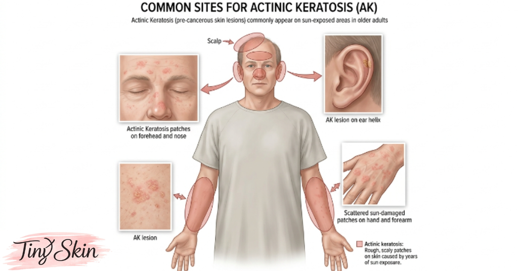
Sun-exposed skin carries the highest risk, so check these areas often.
Face, Ears, Scalp, and Lips
In actinic keratosis pictures, the nose, cheeks, forehead, and tops of the ears appear most often. People with thinning hair also develop patches on the scalp. Ears deserve extra attention because many people forget sunscreen there.
Actinic cheilitis is AK on the lower lip. The lip may look dry, cracked, pale, or scaly, and lip balm does not fix it. Dermatologists take this form seriously because it carries a higher risk of turning into cancer.
Hands, Forearms, and Chest
The backs of the hands and forearms collect years of UV exposure, especially during driving, gardening, or outdoor work. Women may notice pink or tan scaly spots on the upper chest as well. Sunscreen is easy to forget on the hands, which explains why these patches are so common.
How to Check Your Skin at Home
- Stand in bright, natural light.
- Use a mirror and phone camera for hard-to-see spots.
- Photograph any new rough patch next to a ruler or coin.
- Repeat monthly and compare the photos.
Compare your photos with actinic keratosis pictures from trusted medical sources, but never diagnose yourself.
Actinic Keratosis Symptoms: What You See and Feel
Early Signs
Early patches are often easier to feel than to see. Many cause no symptoms at all. Others may bring:
- Roughness like fine sandpaper
- Mild itching or burning
- Tenderness when clothes or jewelry rub
- Dry flaking that returns after you scrub it away
Additionally, the surrounding skin often looks sun-damaged, with freckles, fine lines, or tiny red vessels. Actinic keratosis pictures cannot show roughness, so gently touch the area as well.
Advanced Signs
In advanced actinic keratosis pictures, patches look thicker. They may form a hard, wart-like bump or a cutaneous horn, a cone-shaped growth made of hardened keratin. Others turn darker or bleed after minor rubbing. Thick patches on the ears or lips deserve prompt attention.
Signs of Change That Need Attention
Actinic keratosis can come and go, so a vanished patch is not a cure. However, rapid growth, pain, bleeding, ulceration, or a firm, thick base may signal squamous cell carcinoma. Book a dermatologist visit promptly if you notice any of these changes.
A useful rule: any rough spot on sun-exposed skin that does not heal within a few weeks deserves a professional look.
Types of Actinic Keratosis Compared
Not all actinic keratosis pictures look the same. Dermatologists describe several patterns, and each one affects treatment choices. Knowing these patterns helps you describe your spot clearly at your appointment.
Common Types and Their Appearance
Table 1: Actinic keratosis pictures by type and appearance
| Type | What It Looks Like | Key Note |
|---|---|---|
| Classic (flat) | Pink or red, flat, scaly patch | The most common form |
| Hypertrophic | Thick, rough, wart-like scale | Can resemble squamous cell carcinoma |
| Pigmented | Tan or brown, flat patch | Easily mistaken for a sunspot |
| Cutaneous horn | Hard, cone-shaped growth | May hide cancer cells at its base |
| Actinic cheilitis | Dry, scaly, cracked lower lip | Higher-risk location |
Why the Type Matters
Thin patches may respond to creams. Thick patches often need freezing or a minor procedure. In addition, pigmented and thick lesions may need a biopsy to rule out cancer. Your doctor may also feel the patch, since thickness often matters more than color.
Field Change: The Damage Around the Spot
You may see one patch, yet the nearby skin often carries hidden damage. Dermatologists call this “field cancerization.” Treating the whole area, not just one spot, may lower the chance of new patches. Because of this, a doctor may treat a whole cheek or scalp area, even if only two or three spots are visible.
Actinic Keratosis Pictures vs. Skin Cancer and Look-Alikes
Which Conditions Look Similar?
Several skin problems resemble AK. Actinic keratosis pictures can resemble early skin cancers, so this comparison highlights the main clues.
Table 2: Actinic keratosis pictures compared with look-alike conditions
| Condition | Typical Look | Main Clue |
|---|---|---|
| Seborrheic keratosis | Waxy, brown, “stuck-on” bump | Not precancerous; looks pasted on |
| Squamous cell carcinoma | Firm, scaly, crusted, or ulcerated bump | Grows faster; may bleed easily |
| Basal cell carcinoma | Pearly bump with tiny visible vessels | Shiny surface; sore that will not heal |
| Solar lentigo (sunspot) | Flat, evenly brown spot | Smooth, not rough |
| Eczema or psoriasis | Red, scaly, often itchy patches | Itches more; flares come and go |
How Often Does AK Become Cancer?
Most actinic keratoses never turn into cancer. However, doctors cannot predict which ones will. Research suggests that only a small share progress, yet dermatologists often recommend treating patches instead of just watching them. Squamous cell carcinoma is usually treatable when doctors find it early, which is why prompt checks matter.
When to See a Doctor
Book an appointment if a spot:
- Grows quickly or becomes thick
- Bleeds, oozes, or forms an open sore
- Hurts or itches for no clear reason
- Does not heal within four weeks
- Develops while you take immune-suppressing medicine
Do not wait for pain, because many early skin cancers are painless. Additionally, early checks are quick, and early treatment is usually simpler.
Actinic Keratosis Treatment Options
Actinic keratosis pictures can raise questions, but only an exam leads to treatment. Doctors choose based on the number, thickness, and location of your patches. The goals are simple: remove damaged cells, lower cancer risk, and improve how the skin looks and feels.
In-Office and Prescription Options
Table 3: Common actinic keratosis treatment options
| Treatment | How It Works | Best For | Common Side Effects |
|---|---|---|---|
| Cryotherapy | Freezes the patch with liquid nitrogen | One or a few patches | Stinging, blister, temporary light spot |
| Topical fluorouracil | Cream destroys abnormal cells | Larger areas | Redness, crusting for weeks |
| Imiquimod | Cream boosts local immune response | Face and scalp areas | Redness, flaking, flu-like feeling |
| Diclofenac gel | Anti-inflammatory gel | Mild, thin patches | Dryness, skin irritation |
| Photodynamic therapy | Light-activated medicine | Widespread patches on face or scalp | Burning during light, sun avoidance afterward |
| Curettage | Scrapes off thick patch | Thick lesions | Small wound, scab |
This table is educational only. Your doctor will choose the right option and schedule.
What to Expect During Recovery
Redness, peeling, and crusting are normal with creams and light therapy. Follow your doctor’s schedule exactly, protect the area from sun, and avoid picking the crusts. Call your clinic if you see severe pain, spreading redness, or pus. Healing usually takes a few weeks, and treated skin may look worse before it looks better.
Why Home Remedies Are Risky
Apple cider vinegar, harsh acids, and scrubs may irritate the patch and delay proper care. Moreover, they can hide warning signs. Skip them and see a professional instead.
Prevention: How to Protect Sun-Damaged Skin
Prevention is the best treatment. Actinic keratosis pictures show the price of years of unprotected sun, and that damage adds up over a lifetime.
Daily Sun Protection
Dermatologists recommend these habits:
- Apply broad-spectrum sunscreen with SPF 30 or higher every morning
- Reapply every two hours outdoors, and after swimming or sweating
- Wear a wide-brim hat, sunglasses, and long sleeves
- Use a lip balm with SPF
- Seek shade during midday hours
- Avoid tanning beds completely
Use a generous layer, because most people apply too little. Studies suggest that regular sunscreen use can reduce the number of new patches.
Sun Risks Readers in Russia Should Know
Many people assume they are safe because summers feel short. However, UV exposure adds up. Snow reflects a large share of UV light, so winter sports and long walks matter. Long daylight hours in northern summers also mean more time outdoors. Summer holidays on the Black Sea coast, in Turkey, or in Egypt, plus dacha gardening and outdoor work, bring intense sun exposure. Moreover, tanning salons expose skin to concentrated UV rays.
Regular Skin Checks
Check your skin monthly and see a dermatologist for a full-body exam about once a year, or as your doctor advises. Go sooner if you have many patches, a weak immune system, or a history of skin cancer. Additionally, ask a family member to check your scalp and back.
Key Takeaways
- Actinic keratosis pictures show rough, scaly pink, red, or brown patches, but photos cannot replace a professional diagnosis.
- AK is a precancerous condition caused by long-term UV exposure, and it often appears on the face, ears, scalp, hands, and lips.
- Most patches never become cancer, yet no one can predict which ones will, so early evaluation matters.
- Bleeding, rapid growth, pain, or a thick, hard spot needs a dermatologist visit soon.
- Treatments include cryotherapy, prescription creams, photodynamic therapy, and minor procedures, all chosen by a doctor.
- Daily SPF 30+ sunscreen, protective clothing, and regular skin checks lower your risk of new patches.
FAQs
What do actinic keratosis pictures look like?
Patches usually look small, rough, and scaly, in pink, red, tan, or brown. They often appear on the face, ears, scalp, and hands. Some feel like sandpaper, and thicker ones form a hard, wart-like bump. Because other conditions look similar, a dermatologist should confirm any spot.
Can actinic keratosis go away on its own?
Sometimes a patch fades for a while, especially when you avoid the sun. However, it often returns, and the damaged skin underneath remains. Because doctors cannot predict which patches turn cancerous, they usually recommend treatment rather than waiting. Ask a dermatologist before deciding to monitor a spot.
Is actinic keratosis skin cancer?
Actinic keratosis is not yet skin cancer, but doctors call it precancerous. A small share of patches can develop into squamous cell carcinoma. Since no one can tell which patches will change, dermatologists take every patch seriously. Early treatment removes damaged cells before they become dangerous.
How do dermatologists diagnose actinic keratosis?
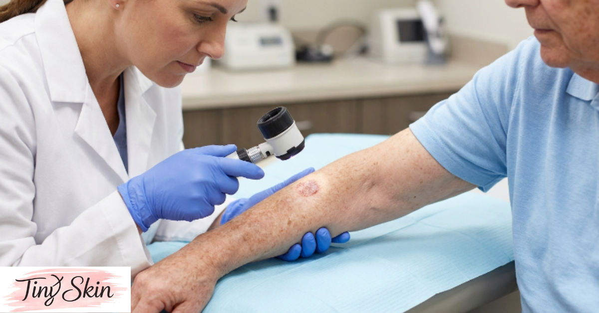
Most diagnoses start with a visual exam and touch. Your dermatologist may use a dermatoscope, a lighted magnifier, to study the patch closely. If the spot looks thick, painful, or unusual, the doctor may take a small biopsy. A lab then checks the cells to rule out skin cancer.
Can I diagnose myself with actinic keratosis pictures?
No. Photos help you notice changes, but lighting, skin tone, and camera quality distort colors and textures. Several harmless and cancerous conditions look similar. Use pictures to prepare questions, then book a professional skin exam. Bring your own dated photos to show how the spot has changed.
Which doctor should I see in Russia for a suspicious skin patch?
Start with a dermatologist (дерматолог) at a public clinic or private medical center. For suspicious or fast-changing lesions, ask for a referral to a dermato-oncologist (дерматоонколог) or an oncology center. Bring your photos, a list of medicines, and details about your sun exposure history.
Conclusion
Actinic keratosis pictures can help you recognize rough, scaly, sun-damaged patches early. However, no photo can tell you whether a spot is harmless, precancerous, or cancerous. Use actinic keratosis pictures as a starting point, not a diagnosis. Only a trained dermatologist can make that call, and early treatment is usually simple.
If you notice a new or changing patch, do not wait. Schedule a skin check with a dermatologist this week. Bring your own photos, and ask which treatment fits your skin. Moreover, protect your skin every day with sunscreen, shade, and regular self-checks. Your skin protects you for life, so return the favor.

